One of the benefits of being a physical therapist (and electromyographer) is the opportunity to spend longer periods of time with our patients, which gives us the chance to ask more questions to try and understand a patient’s symptoms, possible injuries or underlying conditions, to allow us to develop a differential diagnosis of what we could potentially find while performing the nerve conduction studies. Another benefit of having more face time with the patients is that we have the opportunity to answer questions that the patients themselves have about their potential diagnoses and the testing procedures that we will be performing.

As a child, I was frequently taught that “knowledge is power.” That understanding has not only shaped my career as a therapist, but it also gives me the chance to pass along some of my knowledge to patients so that they, too, can increase their own knowledge base and I can help to empower them as they advance through their treatment journey. Patients often come in for electrodiagnostic (EDX) testing with a limited knowledge of what electromyography (EMG) and nerve conduction studies (NCS) are and how they will be of added benefit to understanding their potential diagnoses. So, as it frequently happens, we attempt to break through the medical jargon to explain the testing procedures and the potential diagnoses in a way that is more easily understood.
When a patient is referred for a “nerve conduction study,” there are essentially two parts of the examination that they will experience. First, the nerve conduction study (NCS) utilizes electrical stimulations along the pathway of the nerve to assess the nerve’s health or function. The second part, electromyography (EMG), assesses the health of the muscle and the communication between the nerves and the muscles. If certain abnormalities are present, either in the nerve or in the muscle, our testing can help us identify where the injury is located and to what extent the nerve and/or muscle is affected.
During the nerve conduction study portion of the test, we are assessing each nerve’s responses to electrical stimulations in order to determine if there are any areas where the nerve slows down or if there are any nerves that have less than optimal responses, both of which could indicate nerve compression or damage. We methodically measure out the pathways of the nerve and, with each stimulation, our computers tell us how long it takes for the nerve impulse to travel from where we stimulate to where the responses are being recorded. Therefore, if we know the distance (from our measurements) and we know the time it takes for the nerve to respond, we can calculate the nerve’s conduction velocity, or speed. To explain this a little better, let’s think of our nerves as the roadways connecting different parts of a city and the nerve impulses are the cars travelling along the nerve’s highways. In normal situations, these roadways allow for smooth passage of the cars (nerve impulses) from their starting point, just outside the spinal cord, to their destination, which is either a patch of skin (for sensory nerves) or muscles (for motor nerves). But, as any commuter knows, there can be traffic along the roadways which slows down the ability for the car to continue traveling at a fast or constant speed.
One common diagnosis that we encounter is carpal tunnel syndrome, which causes hand or wrist pain and numbness in the fingers (usually the thumb, index, and middle fingers). The carpal tunnel is a small passageway that the median nerve, along with nine finger flexor tendons, pass through as it enters the hand. This tunnel has a strong ligament that serves as the ‘roof’ and several wrist bones that serve as the ‘floor’ of the tunnel. Imagine that your daily commute has you passing through a tunnel and, when traffic is flowing smoothly, you do not have to alter your speed when passing through. However, if there has been a lane closure within the tunnel, traffic will slow. Similarly, carpal tunnel syndrome occurs when the median nerve, resembling a major highway, faces compression at the wrist’s narrow passageway (the carpal tunnel). By studying and examining the nerve’s traffic flow and speed, we can detect how much more slowly the nerve impulses travel as they cross into the wrist.

Electromyography, or EMG, is the portion of the exam that assesses how well the muscles and nerves communicate. We use a small needle to ‘listen to’ or ‘watch’ the muscle both at rest and with muscle activation. If there is any nerve damage that is affecting the nerve’s ability to reach the muscle, we will see abnormalities on the computer screen, indicating either acute (new/active) or chronic (older/healing) findings. We sample a few different muscles within the arm (or leg) so that we can look for commonalities between the muscles showing abnormalities. For example, if we find abnormalities in two muscles that originate at the same level of the neck (but have different nerves that supply them in the arm), we can be confident that the dysfunction occurs at the neck. Think of the muscles as off ramps on these nerve highways. When nerves transmit signals, muscles respond, much like vehicles exiting the highway and reaching their destination (the muscle). The EMG serves as the traffic camera, capturing muscle responses to nerve signals. By assessing muscle activity both at rest and during activation, we gain insights into the nerve-to-muscle communication, identifying any disruptions or irregularities in the flow of signals.
Another common diagnosis that we see is cervical radiculopathy, or a “pinched nerve” in the neck. While it is common for people to experience neck pain, this does not always imply that the nerve fibers or their insulating structures are damaged. However, when a nerve is affected at the level of the neck, it can lead to pain that radiates into the arm, numbness in specific areas of the arm/hand, and weakness in the muscles that are controlled by the level of the neck that has been affected. Each of the nerves in your arm begins as a nerve root that exits from your spinal cord. As the nerve, or highway, travels throughout the shoulder and into the arm, there are several ‘exit ramps’ that the nerve can take to innervate the different muscles of the arm. For example, your triceps muscle is responsible for extending the elbow and has a specific “exit” along the Radial Nerve “Highway” that originates at the C7 spinal nerve root in your neck.
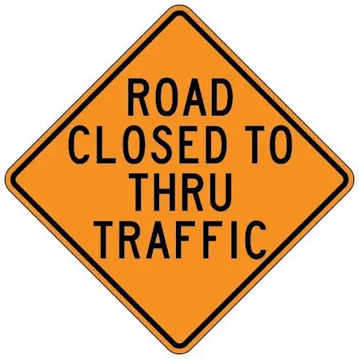
Cervical radiculopathy, or a “pinched nerve” in the neck, is akin to an offramp closure, causing discomfort and pain in the nerve signals that are unable to exit the highway and reach their destination. If there has been damage to the nerve root at C7, the “roadway” of some of the nerve fibers to the triceps muscle (along with other C7 muscles) will be closed which will inhibit “traffic” to that muscle. Since the “traffic” (or nerve signals) is impeded in reaching the triceps muscle, pain and weakness will develop. When a needle is placed within the triceps muscle during EMG testing, there will be abnormal findings that show us how many, if any, of the nerve impulses are still capable of reaching their destination. Identifying the exact level of these “road closures” helps to pinpoint the specific area of injury helping to direct an appropriate treatment plan.
Understanding these tests as traffic analogies simplifies the complexities of nerve conduction studies and electromyography. Just as traffic monitoring ensures smooth transportation, NCV and EMG provide valuable insights into nerve health, enabling healthcare professionals to diagnose nerve-related issues accurately. In essence, while navigating the bustling city of our bodies, these tests act as vigilant traffic cameras, monitoring our neurological highways so that developing problems can be addressed in an appropriate way. So, the next time you hear about nerve conduction studies and EMGs, visualize a busy city with roads, intersections, traffic jams, and road closures – understanding the intricate system that keeps our nerves running smoothly.
Drayton Perkins, PT, DPT


