Electrodiagnostic testing answers a practical question. Is this weakness, numbness, or pain coming from a nerve, a muscle, or the connection between them. Electromyography and nerve conduction studies work together to localize the problem. They also inform prognosis and the next step in care.
This guide explains when a “pinched nerve” appears on testing, when it can be missed, and why timing and muscle selection matter. It also clarifies why sensory studies are often normal in root lesions, and how EMG/NCS complements imaging and clinical history. Physicians and patients can use this to set expectations and plan care.
EMG Solutions delivers board-certified, objective testing. Expert sampling. Same-day interpretation. Clear recommendations to move care forward.
EMG vs NCS, what each test shows
- Needle electromyography (EMG) evaluates muscle electrical activity at rest and with effort. It detects denervation and reinnervation changes from axonal injury.
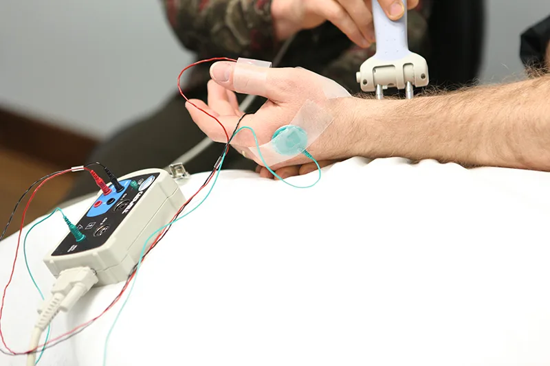
- Nerve conduction studies (NCS) assess sensory and motor nerve signals using surface electrodes and small electrical pulses. They detect demyelination and axonal loss in peripheral segments.
Together, EMG/NCS distinguishes radiculopathy (nerve root) from peripheral entrapment (nerve beyond the root), plexopathy, or primary muscle disease. For a primer on how the tests work, see our overview of the combined electrodiagnostic test.
When a pinched nerve shows up
A pinched nerve shows up when there is axonal injury in a sampled distribution, and when enough time has passed for changes to appear.
- Peripheral entrapments, such as median neuropathy at the wrist or fibular neuropathy at the fibular head, often show abnormal NCS in the involved sensory and motor branches. EMG of muscles innervated by the compressed nerve can show denervation.
- Radiculopathy can show EMG abnormalities in myotomal muscles, including paraspinals. Findings support a root-level problem when two or more muscles from the same root but different peripheral nerves are involved.
Sensitivity improves with targeted muscle selection, adequate sampling, and examiner expertise. Deep paraspinals and key distal muscles increase yield.
When EMG can be normal despite symptoms
A normal study does not rule out nerve pathology. Reasons include timing, lesion type, and anatomical sampling.
- Timing after injury. Fibrillation potentials typically emerge 10 to 21 days after axonal injury. Very early testing can be normal. Distal sensory changes from Wallerian degeneration may lag 10 to 14 days or longer.
- Non-axonal or intermittent lesions. Pure demyelination without conduction block, transient compression, or pain-dominant radiculopathy can produce minimal or no EMG changes.
- Limited sampling. If only a few superficial muscles are tested, a focal lesion can be missed, especially at the root or plexus.
These realities are why EMG/NCS is interpreted with the history, exam, and imaging. A normal EMG does not exclude lumbar radiculopathy.
Radiculopathy vs peripheral entrapment
Radiculopathy originates at the spinal nerve root. Peripheral entrapment occurs along a named nerve distal to the root. This distinction drives expectations on testing.
- Sensory physiology. Sensory nerve cell bodies live in the dorsal root ganglion, which sits outside the spinal canal. In a root lesion proximal to the ganglion, distal sensory responses often remain normal. In a peripheral entrapment distal to the ganglion, sensory responses are typically abnormal in that nerve’s territory.
- Motor findings. Root lesions can denervate muscles from multiple peripheral nerves that share the same myotome. Peripheral entrapment affects muscles downstream of that single nerve.
- Practical implication. Normal sural and superficial fibular sensory studies are common in L5 or S1 radiculopathy, while the same studies may be abnormal if the sciatic or fibular nerve is involved more distally.
Care planning changes when the site of compromise is clear. Imaging and targeted EMG/NCS together improve localization.
Why muscle selection and paraspinals matter
Examiner strategy affects sensitivity. Sampling two or more limb muscles per suspected root, plus the corresponding paraspinals, is recommended. Paraspinal denervation supports a radicular process. Distal limb muscles improve detection of partial lesions. Deep muscles, such as psoas or quadratus lumborum, may be considered when history and exam indicate a higher-level lesion.
EMG Solutions emphasizes precision EMG with expert sampling and same-day reporting to support timely decisions. Learn how an EMG specialist structures studies for clarity on diagnosis and prognosis.
Timing your study after symptom onset
- First 7 to 10 days: EMG may be normal in the limb despite significant pain or sensory change. NCS in radiculopathy often remains normal.
- About 2 to 3 weeks: Denervation potentials typically appear in affected muscles. Sensitivity improves.
- Chronic phase: Reinnervation and chronic neurogenic motor unit changes can clarify severity and chronicity.
If early results are inconclusive but suspicion remains high, repeat testing can be considered. Clinical urgency and evolving deficits guide that decision.
How EMG/NCS fits with imaging and history
Electrodiagnostic testing answers different questions than MRI. MRI shows structure. EMG/NCS shows function. A disc bulge on MRI may be incidental. EMG can help confirm whether a root is physiologically compromised. Conversely, a normal EMG with strong clinical suspicion may still warrant spine imaging or specialist referral. The best care integrates history, physical exam, imaging, and electrodiagnostic data.
What an EMG report includes
An EMG report summarizes history, exam highlights, NCS data, EMG needle findings, interpretation, and clinical correlation. It identifies the most likely site of lesion, severity, chronicity, and differential considerations. It often outlines suggested next steps, such as therapy focus, imaging correlation, or surgical referral when appropriate.
At EMG Solutions, reports are completed the same day to support prompt decision-making. Explore what to expect from an electromyography test and how results are communicated.
Practical next steps after testing
Results guide care, not just labels. Examples include activity modification, targeted physical therapy, splinting for entrapments, anti-inflammatory strategies, injections for selected cases, or surgical consults for severe conduction block or progressive weakness. Coordination with the referring provider remains central.
If your study suggests an ulnar lesion or cervical source of hand symptoms, this educational overview on ulnar nerve vs cervical radiculopathy can help frame the discussion with patients and therapists.
FAQ
- Will a pinched nerve show up on an EMG? Often, yes when there is axonal injury and adequate time has passed. Peripheral entrapments frequently show NCS and EMG changes. Radiculopathy is detected by EMG in myotomal muscles, with sensory studies often normal.
- What is a bad EMG result? There is no single “bad” value. Concerning patterns include active denervation in multiple muscles of a myotome, reduced recruitment, and severe axonal loss or conduction block on NCS. Clinical context determines severity.
- Can you have neuropathy if EMG is normal? Yes. Small-fiber neuropathy often has normal EMG/NCS. Early or purely demyelinating processes can also appear normal. Additional tests or follow-up may be appropriate.
- What is an EMG report? A structured document with history, techniques, objective findings, interpretation, and clinical correlation, used to guide diagnosis and treatment.
- What is the next step after an EMG? Discuss results with the ordering clinician. Typical steps include targeted therapy, ergonomic or activity changes, medications, injections, imaging correlation, or surgical referral if indicated.
Summary and next steps
EMG/NCS is most informative when timed correctly, sampled expertly, and interpreted with the whole clinical picture. Radiculopathy can have normal sensory studies, while peripheral entrapments usually do not. A normal EMG does not exclude a root lesion, especially early after onset. Objective data supports confident decisions and timely care.
For coordinated testing, expert sampling, and same-day reporting, partner with EMG Solutions. Learn more about our EMG specialist services and the combined EMG vs NCS approach, or schedule an electrophysiology consultation to plan the right next step. Internal links:
- Overview of the combined electrodiagnostic test: https://emgsolutions.com/emg-ncs-tests
- EMG specialist services and on-site partnerships: https://emgsolutions.com/emg-specialist-services
- Electrophysiology consultation options: https://emgsolutions.com/electrophysiology-consultation
- Ulnar nerve vs cervical radiculopathy education: https://emgsolutions.com/ulnar-nerve-vs-cervical-radiculopathy/