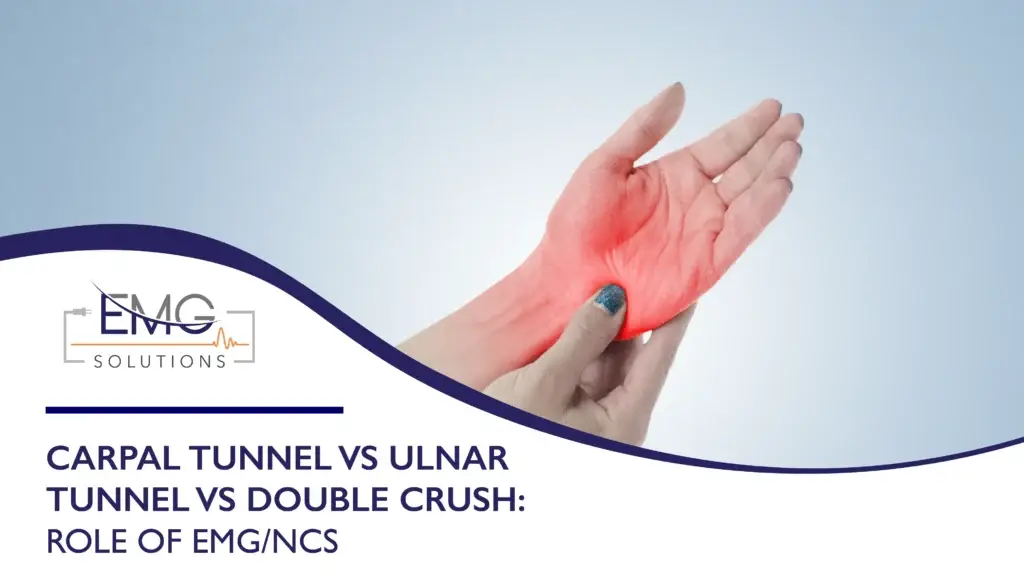
Hand numbness and weakness are common. The source is not always obvious. Median neuropathy at the wrist, ulnar tunnel compromise at Guyon’s canal, or a proximal-plus-distal double crush can look similar in the clinic.
Accurate localization changes treatment. Splinting versus therapy. Injection versus surgery. Timely electrodiagnostic testing clarifies the plan. EMG/NCS provides objective data. Same-day answers keep care moving.
This guide reviews key exam signs, targeted provocative tests, and nerve conduction segments that separate median from ulnar from double crush presentations. It also explains how results inform conservative care and surgical decisions. When you need fast scheduling and same-day reporting, EMG Solutions supports hand surgeons and therapists with on-site testing in partner hospitals and clinics.
Clinical patterns at a glance
Carpal tunnel syndrome, also called median neuropathy at the wrist, typically produces paresthesia in the thumb, index, middle, and radial half of the ring finger. Night symptoms. Shaking the hand out. Thenar fatigue with pinch tasks.
Ulnar tunnel syndrome (Guyon’s canal) can cause palmar sensory symptoms in the small finger and ulnar half of the ring finger, intrinsic hand weakness, or both, depending on which branch is compressed. A lesion of the deep motor branch can cause weakness without sensory loss. Key pinch may be weak. Dorsal ulnar sensation is often spared because the dorsal cutaneous branch leaves proximal to the canal.
Double crush describes the hypothesis that a proximal lesion increases a nerve’s vulnerability to a distal entrapment. The mechanism remains debated, and in practice, the term is often used for coexisting proximal and distal lesions along the same nerve pathway. For the hand, common pairings include cervical radiculopathy with carpal tunnel or cubital tunnel with Guyon’s canal. Symptoms can be broader, with neck or elbow provocation plus distal nocturnal numbness.
Focused examination and provocative maneuvers
History matters. Symptom map. Nocturnal patterns. Neck or elbow pain. Occupational exposure. Diabetes or thyroid disease. Masses or trauma history.
Targeted tests help refine suspicion:
- Median neuropathy: Phalen’s test and Tinel’s at the wrist. Carpal compression test. Thenar strength testing, especially the abductor pollicis brevis.
- Ulnar tunnel: Tinel’s over Guyon’s canal at the wrist crease, ulnar side. Froment’s sign and Wartenberg’s sign suggest ulnar motor involvement. Compare first dorsal interosseous and adductor pollicis strength with hypothenar strength; weakness of the first dorsal interosseous and adductor pollicis with hypothenar sparing suggests a deep motor branch lesion.
- Double crush: Spurling’s for cervical root provocation when dermatomal symptoms or proximal aching exist. Elbow flexion and pressure for the cubital tunnel if ulnar signs appear proximally, while palmar ulnar symptoms persist distally.
Patterns that redirect:
- Dorsal ulnar hand numbness suggests a lesion proximal to Guyon’s canal.
- Thenar atrophy with sensory loss in the median digits supports carpal tunnel. Sensation over the thenar eminence is typically preserved in carpal tunnel syndrome because the palmar cutaneous branch of the median nerve leaves proximal to the carpal tunnel; sensory loss over the thenar eminence points to a median nerve lesion proximal to the wrist.
- Sensory change beyond a single nerve territory, symptoms in the feet, or a length-dependent pattern raises concern for polyneuropathy rather than focal entrapment. Bilateral hand symptoms alone do not, because carpal tunnel syndrome is frequently bilateral.
What nerve conduction studies show and why segments matter
A nerve conduction study (NCS) measures how quickly and effectively signals travel along sensory and motor fibers. It quantifies distal latency, conduction velocity, and amplitude. Segmental testing localizes pathology.
For median neuropathy at the wrist, targeted segments include:
- Median-versus-ulnar sensory comparison to the ring finger, median-versus-radial sensory comparison to the thumb, and median-versus-ulnar palmar mixed nerve comparison.
- Short-segment stimulation across the carpal tunnel to detect focal slowing.
- Median motor distal latency to abductor pollicis brevis.
For ulnar tunnel pathology:
- Ulnar sensory response to digit 5 compared with the dorsal ulnar cutaneous sensory response. An abnormal digit 5 response with a normal dorsal ulnar cutaneous response supports a lesion at or distal to the wrist.
- Motor studies of the first dorsal interosseous and abductor digiti minimi.
- Ulnar motor stimulation across the elbow excludes or identifies a more proximal lesion, and wrist-to-palm stimulation localizes within the wrist and hand when needed.
For suspected double crush:
- Include comparative sensory studies and motor conduction across suspected distal sites, plus needle EMG sampling of myotomes to detect root-level changes. Normal sensory nerve action potentials in nerves not affected by the distal entrapment, together with denervation in muscles that share a root but are supplied by different peripheral nerves, can point toward radiculopathy coexisting with distal entrapment.
What is the normal range for a nerve conduction study?
Normal values vary by lab, limb temperature, age, and nerve tested. Labs maintain reference ranges. As a general concept, distal latencies should not exceed the laboratory’s upper limit of normal, and conduction velocities and amplitudes should not fall below its lower limits of normal. Interpretation is comparative and contextual, not a single universal cutoff.
Needle EMG complements NCS
Needle electromyography (EMG) evaluates muscle activity at rest and with effort. It detects denervation, reinnervation, and recruitment patterns. In median neuropathy, EMG can confirm axonal loss in the abductor pollicis brevis. Because the abductor pollicis brevis is supplied by the C8–T1 roots, evaluating for a coexisting C6–C7 radiculopathy requires sampling C6–C7-innervated muscles such as the pronator teres, flexor carpi radialis, and triceps; a normal study makes radiculopathy less likely but does not exclude it, particularly when symptoms are purely sensory. In ulnar localization, EMG of the first dorsal interosseous and the abductor digiti minimi helps clarify severity and chronicity, and abnormalities in forearm ulnar-innervated muscles, such as the flexor digitorum profundus to the small finger, point to a lesion at or above the elbow rather than at the wrist. In suspected double crush, EMG can identify root-level involvement that nerve conduction studies cannot, although a normal needle examination does not exclude radiculopathy.
Which test is best for nerve damage?
EMG and NCS together. The combined electrodiagnostic test is the standard because each component has strengths that cover the other’s blind spots. EMG/NCS evaluates large-fiber nerve function; it does not assess small-fiber neuropathy, which requires other testing.
How localization guides treatment
Accurate site and severity inform the next step:
- Splinting and activity modification when demyelinating changes are mild without axonal loss.
- Hand therapy for nerve-gliding, posture, ergonomic coaching, and graded strengthening.
- Corticosteroid injection consideration in the carpal tunnel with an appropriate clinical profile.
- Surgical decompression consideration when electrodiagnostic severity is moderate to severe or when weakness and atrophy progress despite conservative care. For a lesion localized to Guyon’s canal, imaging to look for a ganglion or other mass is often warranted.
- For double crush, address both levels. Cervical or cubital contributors plus distal decompression when indicated. Sequence is individualized.
What happens if NCS is abnormal?
Abnormalities guide the plan. Focal slowing suggests demyelination and is generally associated with a more favorable response to decompression or splinting. Reduced amplitudes, particularly with needle EMG evidence of denervation, indicate axonal loss and may signal longer recovery timelines. The electrodiagnostic report should clarify severity and chronicity to support prognosis and follow-up.
Practical tips for clinicians
- Map symptoms carefully, then use two to three targeted provocative tests rather than many unfocused maneuvers.
- Order EMG/NCS when symptoms persist, weakness appears, or surgical planning is on the table.
- Ask for short-segment studies across the wrist when distal entrapment is suspected.
- In a possible double crush, ensure needle EMG includes paraspinals and appropriate myotomes.
For a deeper primer on combined testing and report structure, see an overview of EMG and NCS together as an electrodiagnostic test at EMG Solutions. You can also review how to read a nerve conduction study and the essentials of an EMG report to align your documentation and expectations.
- Learn more about EMG and NCS working together: EMG vs NCS
- Practical guidance on interpreting NCS and EMG reports: Taking the Mystery Out of an EMG/NCS Report – Part 1
Quick FAQ
- How can I tell if my median nerve is damaged?
Look for nocturnal paresthesia in the thumb, index, middle, and radial half of the ring finger, positive carpal compression or Phalen’s, and thenar fatigue. Testing supports the diagnosis when NCS shows focal slowing across the wrist and, when needed, EMG shows axonal loss. Results are interpreted together with the clinical examination, and a normal study does not fully exclude mild carpal tunnel syndrome. - What does a nerve conduction study show?
It measures speed, timing, and size of electrical responses to detect demyelination, axonal loss, or conduction block, and, when focal slowing or conduction block is present, it can localize the lesion to a specific segment. - What is the normal range for a nerve conduction study?
Normal values are lab-specific. Labs control limb temperature during testing and use reference values that account for factors such as age and height. Reports compare your data to reference ranges and side-to-side differences to determine normal versus abnormal. - What happens if NCS is abnormal?
Your care team uses the pattern and severity to guide splinting, therapy, injections, or surgical referral, and to estimate recovery timelines. - Which test is best for nerve damage?
The combination of EMG with NCS is preferred. Together, they improve localization and prognosis.
Fast access to testing and clear reports
EMG Solutions provides on-site electrodiagnostic testing in partner hospitals and clinics. Physical therapists are board-certified in clinical electrophysiology. Reports are usually delivered within 24 hours. Efficient scheduling for hand surgeons and therapists. If you need an EMG specialist or want to understand what the electromyography test includes, explore these resources:
- Find EMG specialist services: EMG Specialist Services
- What to expect from an electromyography test: Electromyography Test
Summary
Median neuropathy at the wrist, ulnar tunnel at Guyon’s canal, and double crush can overlap clinically. Focused exam and provocative tests point the way, but NCS and needle EMG deliver objective localization and severity. Those details shape splinting, therapy, injection, and surgical decisions. When timelines matter, EMG Solutions offers fast scheduling, on-site testing, and reports usually delivered within 24 hours so your hand care plan can move forward with confidence.