The providers at EMG Solutions are always striving to remain at the top of their field while providing ways to help the discipline as a whole. The following was original research performed by one of EMG Solutions’ own providers. This research was presented as a poster presentation at the American Physical Therapy Association’s (APTA) Combined Sections Meeting (CSM).
Surface Anatomy and Nerve Innervation of the Sinus Tarsi: Cadaver Study
Hao (Howe) Liu, *Steve Forbush, *Wen Wang, *Mark Simmons
Physical Therapy Department of University of North Texas Health Science Center, Fort Worth, TX
*Physical Therapy Department of University of Central Arkansas, Conway, AR
Background
Sinus Tarsi Syndrome (STS) is usually diagnosed by clinical symptoms and direct palpation of the area. However, at the time of this research, no anatomical study on cadavers has been done to report the nerve innervations or the surface projection to the sinus tarsi.
Purpose
To measure the surface projections of the sinus tarsi and investigate and describe the nerves which send branches to innervate the sinus tarsi (ST).
Method:
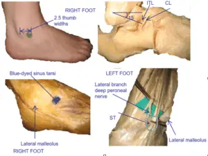
This study was conducted on 20 lower extremities from 10 cadavers (5 male and 5 females ranging from 56 to 86 years at death) with no deformity identified and no scars present in the foot and ankle regions. With the ankle placed at 90 degrees of dorsiflexion, the lateral malleolus (LM) was located and was used as a reference plane (RP). A diagonal line (DL) was placed from the LM to the middle point of the cervical ligament (a landmark structure near the middle of the sinus tarsi) and this line was measured with a digital caliper. The angle between the RP and DL was also measured with a protractor. Fourteen samples from 7 cadavers were dissected to identify the ST surface location, and 6 additional samples from 3 cadavers received injection of a blue dye to verify the results from the dissection study.
Results
The sinus tarsi is a cone-shaped space between the talus and the calcaneus, with its opening toward the dorsolateral side of the hindfoot. The diameter of the opening is around 1.15 cm horizontally. The cervical ligament (CL), along with the interosseous talocalcaneal ligaments (ITL), are thick and strong with the CL lying more superficial and anterolateral to the ITL at the anterior area of the ST. The CL extends vertically from the inferior lateral neck of the talus to the dorsolateral surface of the calcaneus with average length of 1.25 cm. The distance from the LM to the CL is 5.19 cm. The angle between the RP and DL was approximately 15 degrees. The average width of the interphalangeal joints (IPJs) of the thumbs on the dominant side from 43 healthy physical therapy students was approximately 2.0 cm, so the ST is approximately 2.5 thumb widths (5.19/2.00 cm) away from the LM in an anterior inferior direction (about 15 degrees). With these parameters, the verification study using the blue dye injection showed that all fat tissue in the ST was colored blue in all of the samples. The nerve innervations to the ST were studied through delicate dissection. Branches to the ST are mainly from the lateral branch of the deep peroneal nerve (8/14 or 57%), but also were found from the sural nerve (3/14 or 21%), the superficial peroneal nerve (2/14 or 14%), and the tibial nerve (1/14 or 7%).
Conclusions
These results indicate that using the thumb IPJ of an examiner, the surface projection of the ST could be easily located as approximately 2.5 IPJs away from the LM in an anterior interior direction. This space receives innervation primarily from the superficial peroneal nerve but can also be innervated from other nerves branches.
Clinical Significance:
This study presents a method for identifying the approximate location of the sinus tarsi as well as the clarification of possible nerve innervations to the ST. This information may aid clinicians in the examination, differential diagnosis of pain conditions of the lateral foot, and treatment of ankle sprains.